
Prenatal screening and NIPT: a week-by-week calendar of tests in pregnancy
The list of tests handed out at the first visit usually looks frightening. In fact there is a simple logic behind it: in certain weeks of pregnancy certain things can be seen — and not earlier, and not later. A missed “window” often cannot be caught up.
This article is about what to do and when, which tests answer which questions, and how screening differs fundamentally from diagnosis.
Screening and diagnosis are different things
The confusion between these two words causes the most fear, so let us start there.
Screening estimates probability. It answers the question “how high is the risk” and is applied to all pregnant women. A screening result is not a diagnosis. “High risk” means “needs checking further”, not “the baby has the condition”.
Diagnosis gives a “yes” or “no” answer. That requires material from the foetus — chorionic villi or amniotic fluid. Such methods are invasive and carry a small risk, so they are used not for everyone but where indicated.
NIPT, despite its high sensitivity, remains screening. That often surprises patients: a test with over 99% sensitivity for trisomy 21 still needs confirmation by an invasive method before any decisions. The reason is that sensitivity and predictive value are not the same thing: among women with a positive NIPT result, the real share of confirmed trisomy 21 is about 90% in the high-risk group and noticeably less in the low-risk group, and for rarer conditions it is substantially lower.
The calendar of tests, week by week
Up to 10 weeks: the first visit
- confirmation of the pregnancy and an ultrasound to establish the dates, the number of foetuses and the location
- basic tests: a full blood count, blood group and Rh factor, glucose, TSH, ferritin, urinalysis
- HIV, hepatitis B and C, syphilis
- rubella and toxoplasma IgG, if the status is unknown
- a discussion of folic acid and iodine
- blood pressure and BMI — these figures are where the assessment of pre-eclampsia risk begins.
From 10 weeks: NIPT
A non-invasive prenatal test can be done from week 10. It is a test of the mother’s blood, in which cell-free foetal DNA circulates.
What NIPT shows:
- trisomies 21 (Down syndrome), 18 (Edwards syndrome) and 13 (Patau syndrome) — with over 99% sensitivity for trisomy 21
- sex-chromosome abnormalities
- in extended panels — microdeletion syndromes and other conditions. But this is exactly where caution is needed: the clinical value of extended panels is limited, for most rare conditions they give many false positives, and the leading professional societies do not recommend them as routine screening
- the sex of the foetus — a by-product, but a pleasant one for many.
Who benefits most from NIPT: women over 35, a “high” or “intermediate” risk on combined screening, a complicated history, or an anxiety you want to settle without an invasive procedure.
Limitations worth knowing: NIPT does not see structural defects (of the heart, neural tube or limbs) — only ultrasound does that; it is less reliable in multiple pregnancy and with a very high BMI; sometimes the result is inconclusive and the test has to be repeated. And most importantly — a positive NIPT result is confirmed by invasive diagnosis, not acted on directly.
The test is available in the genetic diagnostics section.
11+0 – 13+6 weeks: first-trimester screening
The most important “window” of the whole pregnancy. Missing it means losing the most information obtainable in a single visit.
It has two parts:
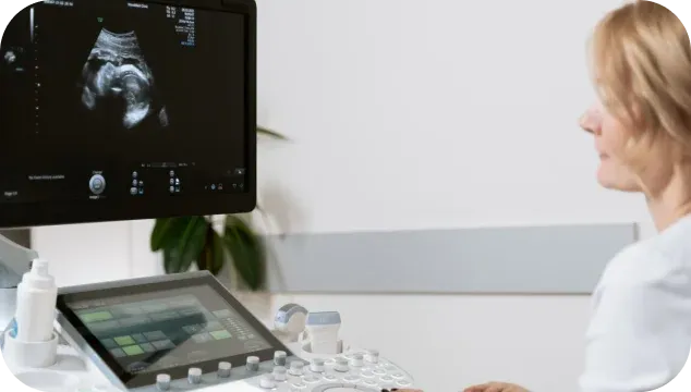
Ultrasound. The crown–rump length is measured (it must be 45–84 mm — which is why the window is so narrow), along with nuchal translucency, the presence of the nasal bone, blood flow, early foetal anatomy, the chorion, and also the length of the cervix and the flow in the uterine arteries.
Blood biochemistry — PAPP-A and free β-hCG.
A computer program combines these data with the mother’s age, weight, gestational age and history, and calculates an individual risk of chromosomal abnormalities. In addition, modern first-trimester screening assesses the risk of pre-eclampsia — and that is perhaps its most practical part: when a high risk is found, prevention started before 16 weeks genuinely lowers the likelihood of severe complications in the second half of pregnancy.
This examination should be done on an expert-class machine by a specialist with the appropriate certificate — here everything depends on the quality of the ultrasound. At Ekstramed this is the expert ultrasound and prenatal diagnostics service.
11–14 weeks: chorionic villus sampling (where indicated)
The first invasive method. Under ultrasound guidance, cells of the chorion are taken and the chromosome set is examined. Indications: a high risk on screening or NIPT, defects on ultrasound, a known genetic condition in the family.
15–20 weeks: second-trimester biochemical screening
Used selectively — for example, if the woman came too late for first-trimester screening. AFP, hCG, free oestriol and sometimes inhibin A are assessed.
From 15–16 weeks: amniocentesis (where indicated)
The standard window is roughly 15–20 weeks; for particular indications the procedure is also done later. A small amount of amniotic fluid is taken with a fine needle under ultrasound guidance. It gives an exact chromosomal and, if needed, molecular-genetic result. By current estimates the risk of pregnancy loss is low — about 0.1–0.3% — which is exactly why the procedure is done only for sound indications, in experienced hands.
Later, after 18–20 weeks, for special indications cordocentesis may be performed — taking blood from the umbilical cord.
18–21 weeks: the foetal anatomy scan
The second key ultrasound. All organs and systems are assessed in detail: the heart with the four-chamber view and outflow tracts, the brain, kidneys, stomach, anterior abdominal wall, spine and limbs. The location of the placenta, the amount of amniotic fluid and the length of the cervix are checked too.
Many structural defects that NIPT cannot see at all are found precisely here. No blood test can replace this examination.
24–28 weeks: the glucose tolerance test
The test for gestational diabetes. Unpleasant but necessary: gestational diabetes often has no symptoms at all, and detecting and controlling it directly affects the course of the pregnancy and the baby’s birth weight. For Rh-negative women, prevention of Rh incompatibility is discussed in the same period.
30–34 weeks: the third ultrasound with Doppler
The growth and weight of the foetus, the state of the placenta, the amount of fluid and the blood flow in the umbilical cord and foetal brain vessels are assessed. The main question at this stage is whether the baby is getting enough nourishment and oxygen.
34–40 weeks: preparing for the birth
Follow-up tests, CTG, assessment of the foetal position and a discussion of the birth plan. Visits become more frequent — every 1–2 weeks.
What to do with a “high risk” result
Calmly and in order:
- Do not read verdicts into the numbers. A risk of 1:100 means that of 100 women with the same result, 99 will not have a baby with this particular chromosomal abnormality. It is heightened vigilance, not a diagnosis
- Consult a geneticist. They will assess the result in the context of your history, ultrasound and age — and tell you exactly what to check next
- Choose the next step: NIPT as a refining screen, or invasive diagnosis as the final answer. The choice depends on what raised the suspicion: with defects on ultrasound it is usually an invasive test, with a “numerical” risk and no findings NIPT is an option
- Do not rush into decisions before a confirmed diagnosis. False-positive screening results are an everyday occurrence — that is what the second stage of screening is for.
Frequently asked questions
Can first-trimester screening be replaced by NIPT? No, they are not interchangeable. NIPT is more accurate for trisomies, but it does not assess foetal anatomy, cervical length or the risk of pre-eclampsia. The optimum is an ultrasound at 11–13 weeks combined with NIPT or combined screening.
Is NIPT safe? Yes. It is an ordinary blood draw from the mother’s vein; there is no risk to the foetus at all.
What if I first saw a doctor at 15 weeks? First-trimester screening is no longer possible, but NIPT, second-trimester biochemical screening and a full anatomy scan at 18–21 weeks are available. Come in without putting it off any further.
Is amniocentesis compulsory after a bad NIPT? Before any decisions the diagnosis has to be confirmed. NIPT is screening, and false positives happen, especially for rarer conditions.
How many ultrasounds can be done in a pregnancy? Diagnostic ultrasound has no proven harmful effect on the foetus. The number is determined by medical indications, not by a “limit”.
Where to have prenatal screening in Ivano-Frankivsk? The Ekstramed clinic offers expert ultrasound, combined screening, NIPT and invasive diagnosis, as well as pregnancy care programmes with every test at the right time. To book — the contacts section.
This article is for information only and does not replace a consultation with a doctor. The scope and timing of tests are decided individually by an obstetrician-gynaecologist.
More worth reading
More explanations, advice and answers from the doctors of the clinic.
 01/09/2026Technology9 min readHow IVF works: every stage of the programme, step by step
 01/09/2026Preparation7 min readPreparing for pregnancy: which tests a couple should have before conceiving
 01/09/2026Technology7 min readSemen analysis: how to read the result and what to do next
 01/09/2026Technology7 min readEgg donation: when it is needed, how a donor is chosen and what the chances are
- Technology01/09/20269 min readHow IVF works: every stage of the programme, step by step
- Preparation01/09/20267 min readPreparing for pregnancy: which tests a couple should have before conceiving
- Technology01/09/20267 min readSemen analysis: how to read the result and what to do next
- Technology01/09/20267 min readEgg donation: when it is needed, how a donor is chosen and what the chances are