
Congenital uterine anomalies: why an accurate diagnosis needs 3D ultrasound
A woman can go for years without knowing that her uterus has an unusual shape, until she faces a miscarriage, a premature birth or repeated failed attempts to conceive. The cause may be a congenital uterine anomaly, also called a Müllerian anomaly. These are not rare: according to current data, 4–7% of all women have one, and among patients with recurrent pregnancy loss the figure is 13–25%.
This article is based on a talk by Andrii Sukhotskyi, ultrasound doctor at the Ekstramed clinic, entitled “Müllerian anomalies: the evolution of diagnosis from 2D to 3D”. We explain what kinds of anomaly exist, why they matter and why a standard ultrasound is often not enough for an accurate diagnosis.
Where uterine anomalies come from
The uterus forms before birth from two paired tubes, the Müllerian (paramesonephric) ducts. They give rise to the uterus, the fallopian tubes, the cervix and the upper part of the vagina. The key events happen in two stages:
- at around the 10th week of foetal development the ducts fuse into one. If they fail to fuse, or fuse only partly, the result is a unicornuate, bicornuate or double uterus;
- at around the 20th week the wall between the fused ducts is reabsorbed. If it is not reabsorbed completely, a septum remains in the uterus, or the top of the uterus keeps a saddle-shaped (arcuate) dip.
A separate group consists of errors in organ formation itself, when the uterus does not form at all or remains rudimentary. Vaginal septa also belong here: a transverse septum can block the outflow of menstrual blood, while a longitudinal one often occurs together with a bicornuate or double uterus.
Important: about 30% of women with uterine anomalies also have anomalies of the urinary tract, such as a missing or unusually positioned kidney. The genital and urinary tracts share a common embryonic origin, so whenever a uterine anomaly is found, the kidneys are always examined too.
How uterine anomalies show themselves
Outside pregnancy, a woman may see a doctor because of:
- no periods from the start of puberty (primary amenorrhoea);
- very painful periods or pelvic pain;
- infertility, as an unusually shaped cavity lowers the chance of embryo implantation;
- a mass in the vagina when menstrual blood cannot drain;
- associated anomalies of the kidneys or skeleton.
During pregnancy the list of possible complications is longer:
- miscarriage and recurrent miscarriage: a uterine septum raises this risk to as much as 44%;
- premature birth: up to 20% with a bicornuate uterus;
- the waters breaking too early, and restricted foetal growth;
- abnormal placental attachment, and bleeding before and after birth;
- cervical insufficiency, an abnormal position of the baby, and caesarean section;
- most dangerous of all, rupture of a rudimentary horn if the pregnancy develops inside it.
That is why early and accurate diagnosis of a uterine anomaly is not an academic exercise but a practical necessity: it determines how a pregnancy is managed and whether surgery is needed.
Why a standard ultrasound is not enough
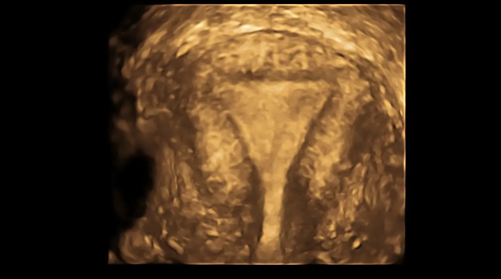
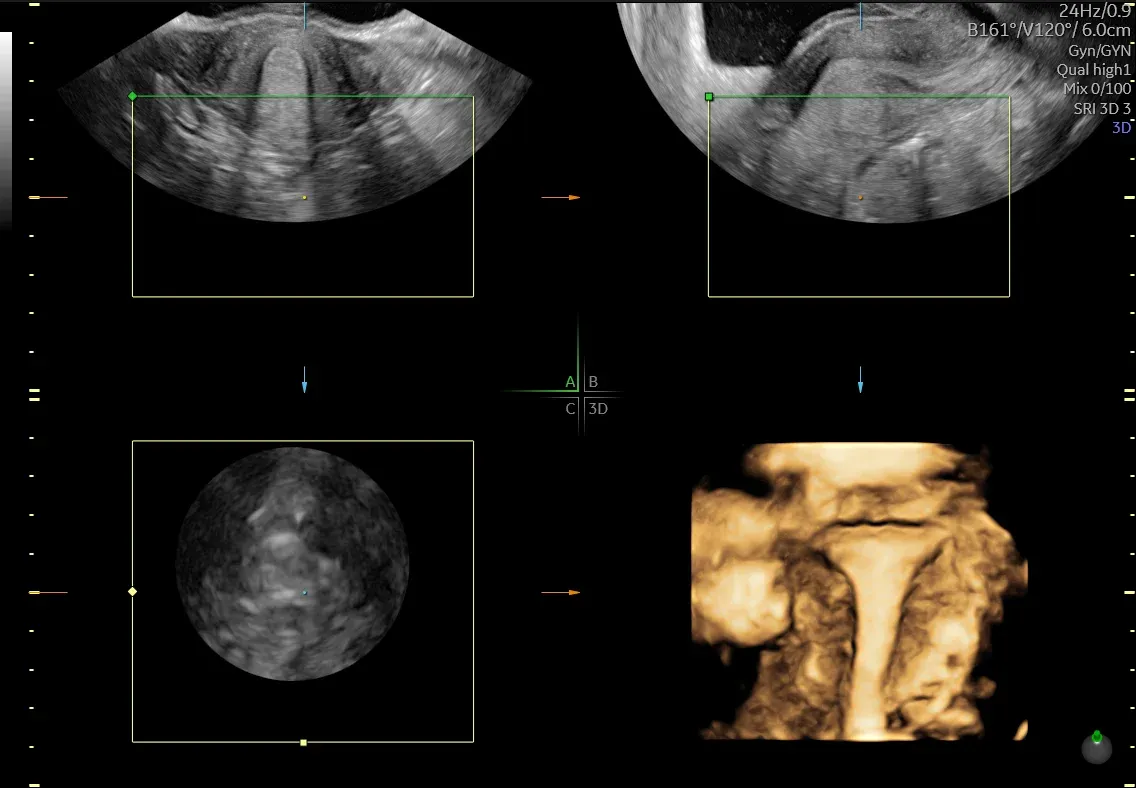
On a standard 2D ultrasound the doctor sees the uterus in longitudinal and transverse sections. But the most informative view for assessing the shape of the cavity is a different one, the coronal (frontal) plane. Only on this plane can the doctor see at the same time:
- the outer contour of the top of the uterus (the fundus);
- the inner contour of the cavity;
- the openings of both fallopian tubes.
This is where the depth of the external fundal indentation, the internal indentation and the angle between the horns are measured, and these figures decide the diagnosis. The problem is that with the uterus in its usual position, obtaining this plane on a 2D machine is technically difficult or impossible. This is where three-dimensional (3D) ultrasound comes in.
What 3D ultrasound offers
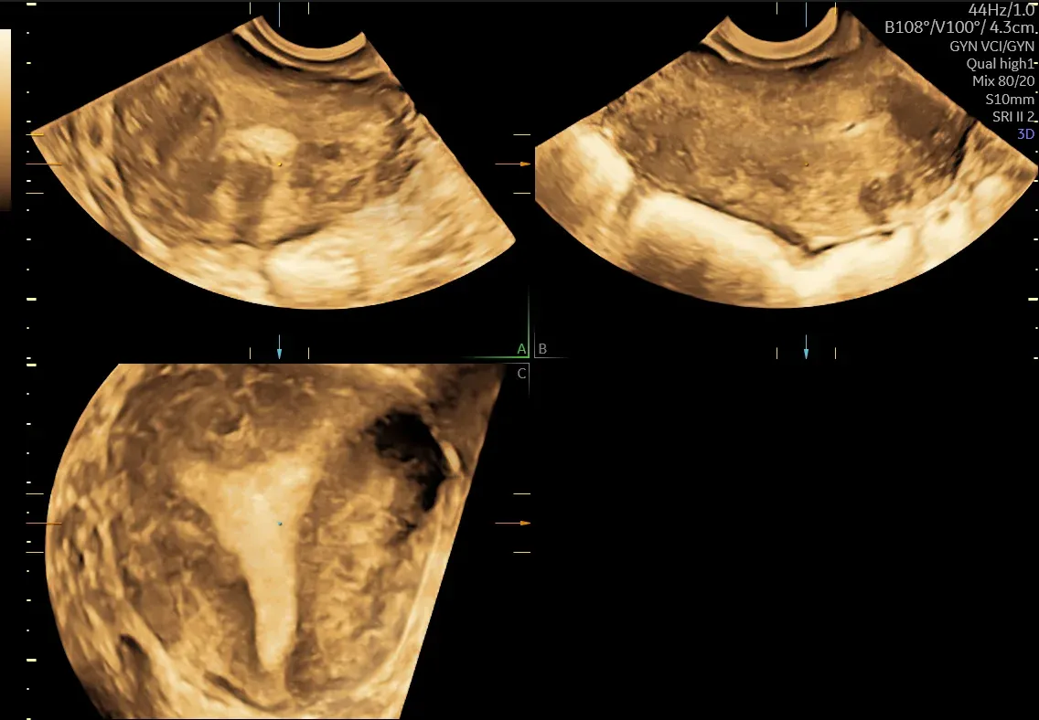
During a 3D scan the machine records the whole volume of the uterus, from which any section can then be “cut”. GE Voluson ultrasound systems have several tools for this:
- multiplanar mode: three mutually perpendicular planes from a single volume, and the main way to obtain the coronal plane “in one click”;
- VCI (Volume Contrast Imaging): adjusts the slice thickness and improves contrast, which is especially valuable for assessing the myometrium and septa;
- OmniView: a section along any line or curve, useful for a uterus of unusual shape or one tilted backwards (retroflexed);
- TUI: up to eight parallel slices at once, so the uterus can be “leafed through” layer by layer, as on CT or MRI;
- VOCAL: measures volume, of a rudimentary horn for example, and assesses blood flow with 3D Doppler;
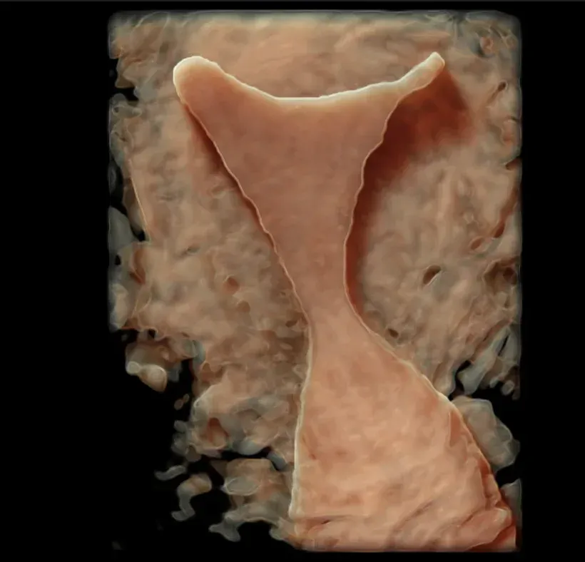
- HDlive: a realistic volume image with virtual lighting, clear for documentation and for explaining findings to the patient.
According to a meta-analysis (Graupera et al., 2015), the sensitivity and specificity of 3D ultrasound in diagnosing uterine anomalies are comparable to those of MRI and exceed 90%. Standard 2D ultrasound performs considerably worse, especially when a septate uterus has to be told apart from a bicornuate one.
When to have the scan
The best time is the secretory phase of the cycle, days 21–24, when the endometrium is at its thickest and stands out well against the muscular wall. The doctor assesses the outer contour of the fundus, the shape of the cavity, whether there is a septum, the angle between the horns and the ratio of the external to the internal indentation depth, which is the key to the diagnosis.
The main types of anomaly
Normal and arcuate uterus
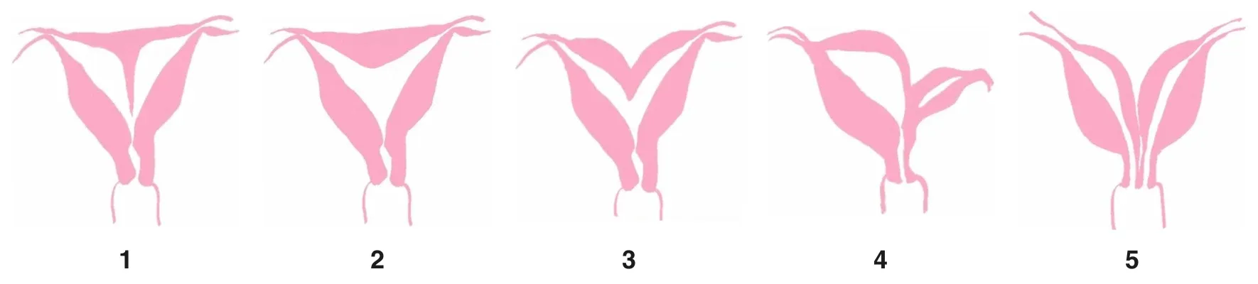
An arcuate (saddle-shaped) uterus is a borderline variant of normal: the outer contour is smooth or has an indentation of up to 10 mm, and the cavity has a slight curved dip at the top (up to 10–15 mm). Under the European ESHRE/ESGE classification it is a variant of normal (class U0), whereas the American ASRM classification places it in a separate class VI. As a result, the same report may be interpreted differently by doctors working with different systems.
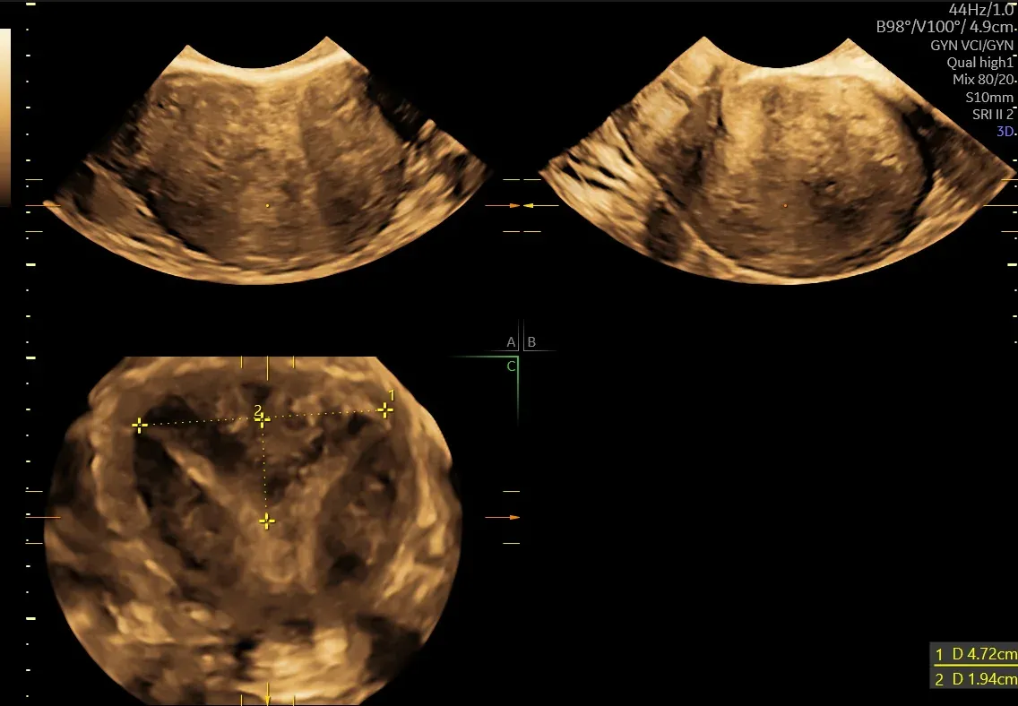
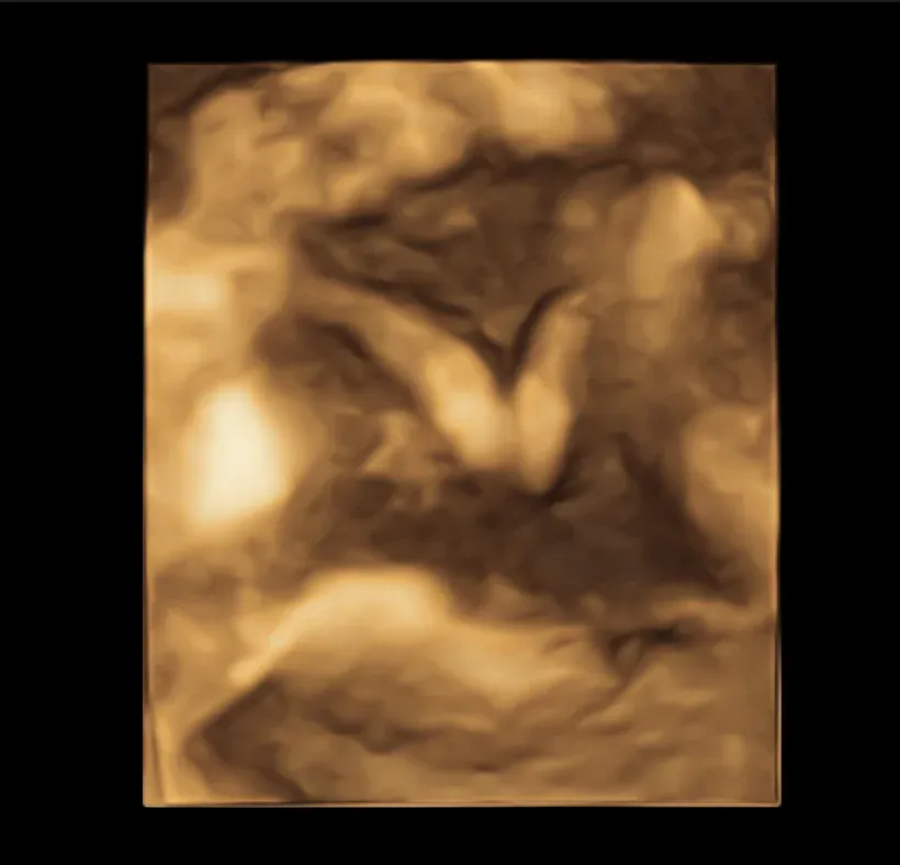
Septate uterus (U2)
The most common anomaly, accounting for up to 35% of all cases. The outer contour of the uterus is normal, but inside a septum divides the cavity, either partly (U2a) or completely, down to the level of the internal os (U2b). The septum is the anomaly most often linked to miscarriage, and it is also the one that can be removed by hysteroscopy, a minimally invasive operation performed through the cervix.
Bicornuate uterus (U3)
Here the body of the uterus is split in two: the outer contour of the fundus has an indentation of more than 1 cm (or more than 50% of the wall thickness), and two muscular horns, each with its own cavity, join above the cervix. There is usually a single cervix and vagina. Partial (U3a), complete (U3b) and combined forms with an additional septum (U3c) are distinguished. Surgical correction of a bicornuate uterus is much more complex, the indications for it are limited, and in most cases the pregnancy is managed conservatively.
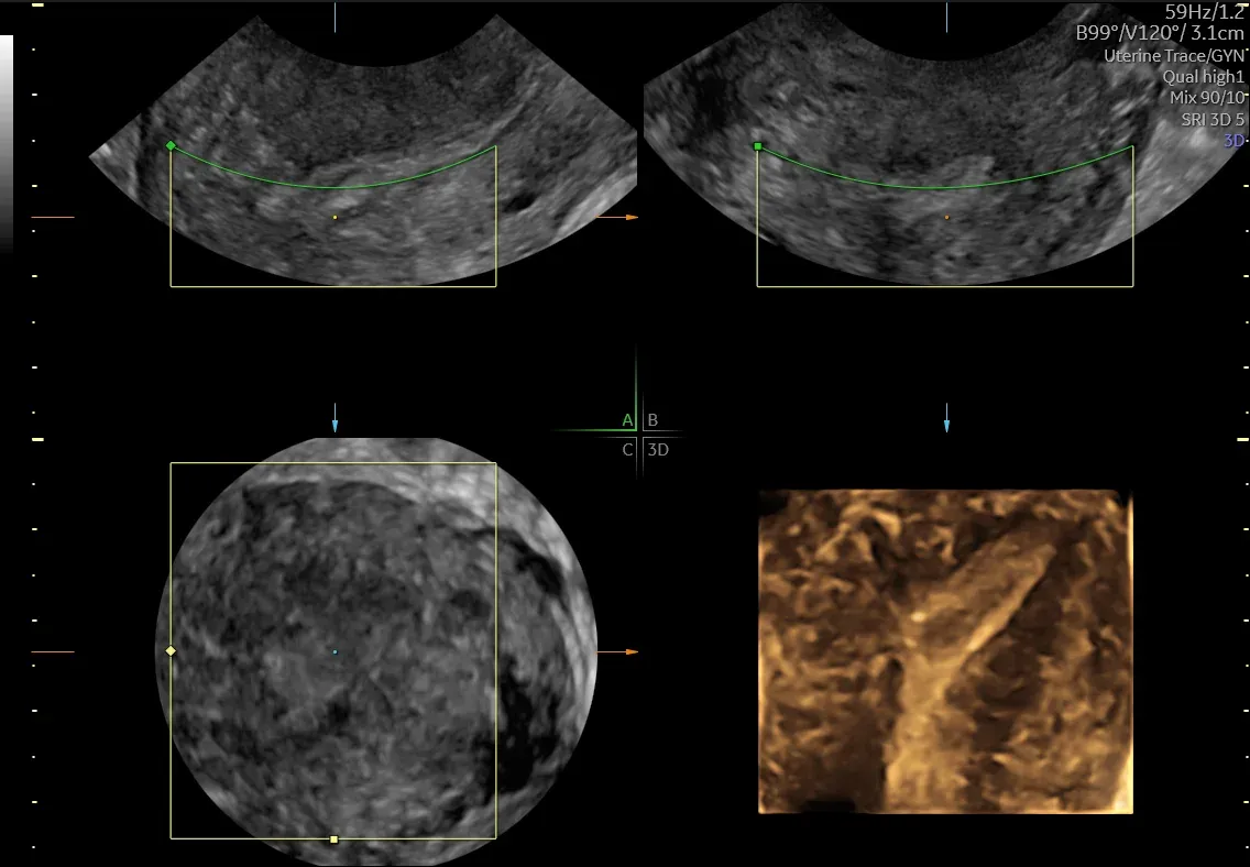
Unicornuate uterus, or hemi-uterus (U4)
Only one Müllerian duct develops; this accounts for about 20% of uterine anomalies. The other half may be absent or remain as a rudimentary horn:
- with a functional cavity (U4a): the most dangerous variant. Menstrual blood from the horn cannot drain, which leads to endometriosis and pelvic pain. If a pregnancy develops in the rudimentary horn, it may rupture at 10–20 weeks with massive haemorrhage. For this reason such a horn is removed laparoscopically;
- without a functional cavity (U4b): the horn can mimic a fibroid, which the doctor bears in mind when making a differential diagnosis.
Uterus didelphys
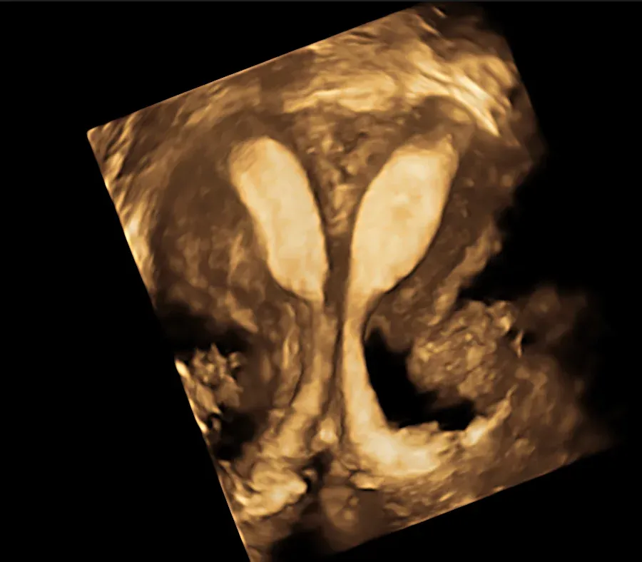
The ducts do not fuse at all: two separate uteri form, each with its own cervix, often with a longitudinal septum in the vagina. The anomaly often causes no symptoms; the septum may cause pain during sex or difficulty using tampons. The two uterine bodies are set wide apart, so here, unlike with other anomalies, diagnosis is often actually better on 2D. Variants in which half of the vagina is closed off need urgent treatment: division of the septum.
T-shaped uterus (U1) and MRKH syndrome (U5)
A T-shaped (dysmorphic) uterus has a normal outer contour but a narrow cavity because of thickened side walls. In Mayer–Rokitansky–Küster–Hauser (MRKH) syndrome the uterus and upper vagina are absent or only rudimentary, although the ovaries work normally, the karyotype is female (46,XX) and puberty happens on time. The main complaint is the absence of periods. Because the eggs do mature, IVF with surrogacy is an option for these women.
Septate or bicornuate: the key question
The most important diagnostic task is to tell a septate uterus apart from a bicornuate one. This decides whether the patient is referred for hysteroscopic division of the septum or monitoring is chosen. A wrong diagnosis means the wrong treatment.
The cavity can look similar in both cases, and the difference shows only on the outer contour, that is, on the coronal 3D plane. Different classifications draw the line in different places:
- ASRM (American, updated in 2021): a septate uterus has a smooth outer contour (an indentation of less than 1 cm), an internal indentation of more than 1.5 cm and an angle of less than 90°; a bicornuate uterus has an external indentation of more than 1 cm;
- ESHRE/ESGE (European, 2013): measures the indentation not in millimetres but as a percentage of the uterine wall thickness; a uterus is septate when the internal indentation exceeds 50% of the wall thickness and the external one is less than 50%;
- CUME 2018: a consensus that brought together the views of 769 specialists from 56 countries. It defines a septum that needs surgery as follows: an internal indentation of more than 10 mm, an angle of less than 140°, and a ratio of indentation depth to wall thickness of more than 110%.
In the speaker’s view, ESHRE tends towards overdiagnosis and unnecessary operations, ASRM can lead to a problem being overlooked, and the CUME 2018 criteria give the most accurate and safest guide. That is why it matters for the report to state not only the name of the anomaly but also the measured depths and angles.
Key points
- Congenital uterine anomalies affect 4–7% of women; they influence both the ability to conceive and the course of pregnancy.
- An accurate diagnosis needs a coronal plane of the uterus, and 3D ultrasound is the method of choice for obtaining it.
- The best time for the scan is days 21–24 of the cycle.
- When a uterine anomaly is found, the kidneys are examined as well.
- Most importantly, a septum that can be removed hysteroscopically has to be told apart from a bicornuate uterus.
If you have had miscarriages, a premature birth or infertility, book a 3D ultrasound of the uterus or an appointment with a gynaecologist at Ekstramed in Ivano-Frankivsk.
This article is for information only and does not replace a consultation with a doctor. The diagnosis and treatment are decided by a doctor after an examination.
Sources: talk by A. Sukhotskyi, “Müllerian anomalies: the evolution of diagnosis from 2D to 3D”; AFS/ASRM (1988), ESHRE/ESGE (2013) and ASRM Müllerian Anomalies Classification 2021; Ludwin A. et al., Congenital Uterine Malformation by Experts (CUME), 2018; Graupera B. et al., 2015.
More worth reading
More explanations, advice and answers from the doctors of the clinic.
 01/09/2026Technology7 min readPrenatal screening and NIPT: a week-by-week calendar of tests in pregnancy
 01/09/2026Preparation7 min readPreparing for pregnancy: which tests a couple should have before conceiving
 01/09/2026Technology9 min readHow IVF works: every stage of the programme, step by step
 18/11/2026Technology6 min readThe Applebaum score: how ultrasound and Doppler show whether the uterus is ready for embryo transfer
- Technology01/09/20267 min readPrenatal screening and NIPT: a week-by-week calendar of tests in pregnancy
- Preparation01/09/20267 min readPreparing for pregnancy: which tests a couple should have before conceiving
- Technology01/09/20269 min readHow IVF works: every stage of the programme, step by step
- Technology18/11/20266 min readThe Applebaum score: how ultrasound and Doppler show whether the uterus is ready for embryo transfer